Technical Data Sheet
ROTI®Lumin, 2 x 120 ml
2 x 120 ml is sufficient for approx. 3,500 cm2 membrane (approx. 50 mini gel blots) or 2,400 wells in the ELISA.
€504.20/Pack Qty.
excl. VAT. | 1 kit per Pack Qty.
Art. No. P078.1
Alternative products

Fits to / Accessories


Product details
- Sensitive and reliable
- For all standard membrane types
- Low background
- No cumbersome preparation of peroxidase buffers
- Suitable for multiple stripping and hybridisation
ROTI®Lumin is a kit for chemoluminescent detection of peroxidase-linked reporters (e.g. HRP-antibodies), as can be found in Western-Blot assays. In comparison to chromogen substrates, ROTI®Lumin displays increased sensitivity and reduced background reaction.
ROTI®Lumin is transformed by the HRP into an excited dianion in the presence of hydrogen peroxide. When the dianion is reversed back to its normal state, light is emitted. The light emission of ROTI®Lumin reaches its maximum within 5 minutes and remains at the maximum level for approx. 1-2 hours.
ROTI®Lumin 10x conc., fluorescence-free, for protein and cytoimmunochemistry
ROTI®Lumin consists of two solutions, which should be mixed at a 1:1 ratio before use. It can be used with nitrocellulose, nylon and PVDF membranes. Optimal results can be achieved with Immobilon-P® PVDF or ROTI®PVDF.
Can be used for Western-Blotting, ELISA, Southern- Blotting, Dot-Blotting and colony hybridisation. Membranes may be stripped from bound probes using ROTI®Free stripping buffer, and may thus undergo multiple reprobing.
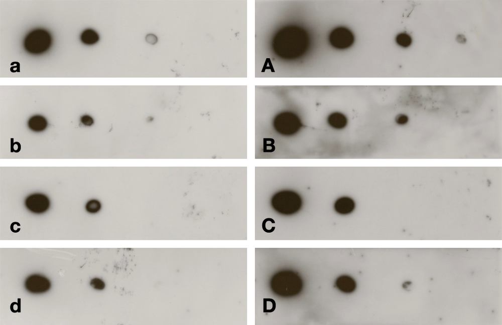
Sensitivity of ROTI®Lumin ultra is found in low femtogram range. Detection of a polyclonal antibody (left to right 4 pg, 400 fg, 40 fg, 4 fg) on ROTI®PVDF (Art. No. T830.1). Staining in PBST via a HRP-conjugated anti-rabbit antibody (1:2000).
a/A: ROTI®Lumin ultra; b/B: ROTI®Lumin plus; c/C: ROTI®Lumin; d/D: Product of a competitor, recommended for femtogram range. a/b/c/d: 10 sec. exposition, A/B/C/D: 1 min. exposition.

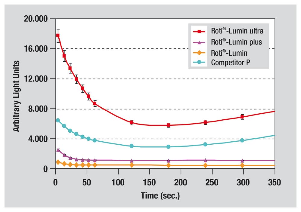
Typical course of light emission of the ROTI®Lumins: 0-30 mins., post mixing/spraying. Competitor P: Product of a competitor, recommended for femtogram range.
120 ml (25 ml) ROTI®Lumin 1 (Art. No. P079) and 120 ml (25 ml) ROTI®Lumin 2 (Art. No. P080).
Contents of this Kit may not be bought separately.

Relative sensitivity of ROTI®Lumins. Measurement of the relative light emission following ECL reaction with solubilised HRP.
Competitor P: Product of a competitor, recommended for femtogram range.
Typical course of light emission of the ROTI®Lumins: 0-5 mins., post mixing/spraying. Competitor P: Product of a competitor, recommended for femtogram range.
| Application | Western, in situ, Histochemistry |
| Enzyme | Horseradish Peroxidase |
| Detection | chemoluminescent |
| Signal product | Precipitate (Blot) |
| Use | Western |
- Subtotal: 0.00
| Art. No. | Pack Qty. | Pack. | Packaging | Price | Quantity | |
|---|---|---|---|---|---|---|
| P078.1 | 1 kit | plastic | 2 x 120 ml |
€504.20 |
|
|
| P078.2 | 1 kit | plastic | 2 x 25 ml |
€134.40 |
|
|
|
In stock
Available
In procurement
No longer available
Delivery date currently unknown
|
||||||
- Subtotal: 0.00
Downloads / MSDS
General information
Stock solutions (All solutions are stable for at least 1 year at 4 °C):
0.5 g NBT in 10 ml 70 % dimethylformamide;
0.5 g BCIP (p-toluidine salt) in 10 ml 100 % dimethylformamide.
Incubation buffer for alkaline phosphatase:
100 mM NaCl, 5 mM MgCl2,100 mM Tris (pH 9.5).
Fresh substrate solution: 66 μl NBT stock solution + 10 ml incubation buffer, mix well, add 33 μl BCIP stock solution. Use within 1 hour.
Blot development: Approx. 10 ml substrate solution per 15 x 15 cm2 membrane surface. Develop at room temperature until bands become visible (approx. 30 mins).
Reaction stop: Rinse with PBS/20 mM EDTA.
Dissolve 1 mg TMB in 0.1 ml dimethylsulfoxide (Art. No. 4720); add 9.9 ml of a 0.1 M sodium acetate solution (pH 6.0) (Powder: Art. No. 6779), filter and add H2O2 (Art. No. 8070) (final concentration 0.01 %).
Always prepare freshly!
Incubation 10-30 mins at room temperature (approx. 50 μl per microtitre well; finally add 50 μl 1 M H2SO4, Art. No. X873, per well).
Photometric quantitation at 450 nm.
Reference: Bos E.S. et al. (1981) J. Immunoassay 2, (3/4), 187.